A pathologist can only diagnose certain masses from an intact piece of tissue, and that is the reason a biopsy sometimes wins out over the faster, gentler fine needle aspirate. It is tempting to believe an experienced veterinarian can tell a harmless fatty deposit from cancer by feel, but no one can, which makes sampling the one dependable way to know what a lump actually is. An aspirate draws a scattering of cells through a needle not much bigger than the one used for a vaccine, and for plenty of skin lumps in dogs and cats, that small sample settles the question. When the cells come back inconclusive, or when the mass sits somewhere that makes treatment planning hinge on precise detail, stepping up to a biopsy is what gets your pet a genuine diagnosis.
Creature Comforts Veterinary Service in Saylorsburg starts many lump conversations with VISTA iQ lump and bump cancer screening, a non-invasive heat-imaging scan that helps flag which of a dog’s skin masses deserve a closer look. When a mass needs tissue, we can guide the biopsy with ultrasound for precise placement, and our in-house lab runs the supporting bloodwork before any procedure. A new lump is easier to deal with when you act early, so give us a call and we will help you choose the test that fits your pet.
Needle Test or Biopsy: The Basics
- No one, not even an experienced veterinarian, can tell a benign lump from cancer by look or feel alone, so sampling is the only reliable way to know what a mass is.
- A no-needle screening scan can sort a dog’s lumps by cancer risk in minutes, a fine needle aspirate gives a gentle first diagnosis, and a biopsy answers what cytology can’t, like a tumor’s grade and margins.
- Some problems, such as splenic masses and chronic intestinal disease in cats, skip the needle test entirely because tissue architecture is the only path to a dependable diagnosis.
- Catching a lump early gives your pet more options, whether the answer turns out to be reassuring or serious.
Can a Vet Tell if a Lump Is Cancer Just by Looking at It?
No veterinarian, however experienced, can reliably tell a benign lump from a cancerous one by sight or touch alone. A soft, movable mass might be a harmless fatty deposit or an early soft tissue sarcoma, and the two can feel exactly the same. Cancer in pets is far more common than most pet families expect, developing in close to one in four dogs at some point and growing more likely as pets age.
Several skin masses in dogs and cats look nearly identical from the outside yet carry completely different prognoses.
Which Skin Lumps Show Up Most Often on Dogs?
Dogs grow a whole catalog of bumps over a lifetime, and most of them are benign.
- Histiocytomas and cysts often look alarming but usually aren’t. A histiocytoma is a benign, button-shaped growth that often appears suddenly on a younger dog and can shrink away on its own within a couple of months. Cysts are fluid- or debris-filled pockets that feel firm and can rupture or refill.
- Mast cell tumors are the great impersonators. Mast cell tumors are one of the most common malignant skin cancers in dogs, and they’re notorious for disguising themselves as an ordinary wart or fatty lump.
- Lipomas feel reassuring, but not every soft lump is one. Lipomas in dogs are soft, benign fatty deposits common in middle-aged and older pets, yet a soft tissue sarcoma or mast cell tumor can feel almost identical to the touch.
- Melanocytic tumors range from harmless to serious. Pigmented, dark masses on the skin, nails, or in the mouth can be simple moles or true melanomas, and only sampling separates the two.
What Lumps Are Most Common on Cats?
Cats grow fewer skin lumps than dogs, but a larger share of feline masses turn out to be malignant, and cats are experts at hiding discomfort. By the time a bump gets noticed, it may already be advanced.
- Basal cell tumors lean benign. These common feline skin nodules are usually slow-growing, though a small portion turn out to be malignant and need removal.
- Squamous cell carcinoma targets sun-exposed skin. Squamous cell carcinoma in cats is an aggressive cancer that favors thinly haired, sun-exposed spots like the ears, nose, and eyelids, and it’s often advanced by the time it’s spotted.
- Fibrosarcomas run firm, deep, and invasive. These connective-tissue growths tend to anchor into the layers beneath the skin and return if any is left behind.
- Mast cell tumors appear in cats too. Feline mast cell tumors rank among the most common skin tumors in cats and frequently show up as small, firm, raised nodules that are easy to brush off.
Is There a Way to Screen My Dog’s Lump Without a Needle?
Yes. Our VISTA iQ lump and bump screening assesses a dog’s lump in real time with no needles at all: a 40-second scan, results in about two minutes, and a cancer risk level that guides what happens next. It’s a screening step rather than a final diagnosis, and its job is telling us which lumps need sampling and which can simply be watched.
The technology, called Heat Diffusion Imaging, gently warms the lump by a few degrees and then measures how the tissue warms and cools. Healthy and unhealthy tissue heat and cool at different rates because their blood supply, metabolism, and structure differ, and a trained, validated artificial intelligence system reads that pattern.
A low-risk result usually means we watch the lump for changes. A moderate or high-risk result starts a conversation about sampling, which is exactly where the needle test and biopsy below come in. One honest limit: the VISTA iQ is designed specifically for dogs, so for a lump on your cat, the fine needle aspirate remains the first step.
What Is a Fine Needle Aspirate, and Does It Hurt?
Fine needle aspiration collects a small sample of cells through a thin needle, takes only a few minutes, usually needs no sedation, and typically returns answers within a few business days. The whole thing is over so quickly that most dogs and cats tolerate it with little more than a wiggle.
Once the sample is on a slide, a pathologist studies the cells under a microscope, judging their shape, size, and how they cluster to sort them into categories like fatty, inflammatory, or cancerous. When a mass sits deeper, near an organ or inside the belly, we use ultrasound guidance to steer the needle precisely into the target.
Cytology shines with certain tumors: lipomas, many mast cell tumors, and abscesses often give a clear result from a single aspirate. Its limit is that it only shows loose, individual cells, not how they’re arranged. Some masses don’t shed cells well onto a slide, and others need the surrounding architecture to be graded. When the sample comes back inconclusive, or too thin to read, a biopsy is the logical next step.
When Does a Pet Need a Biopsy Instead of an Aspirate?
A biopsy becomes necessary when the diagnosis depends on tissue structure rather than loose cells. Cytology versus biopsy comes down to reading individual cells against reading intact tissue, and only tissue can reveal a tumor’s grade, how deeply it has invaded, and whether the surgical margins came back clean.
Histopathology, the study of that intact tissue, tells us not only what a tumor is but how it’s likely to behave. Grade is a score for how aggressive the cells look, and it helps predict whether a cancer tends to spread. Margins describe the rim of normal tissue around a removed mass, and clean margins mean the surgeon very likely got all of it.
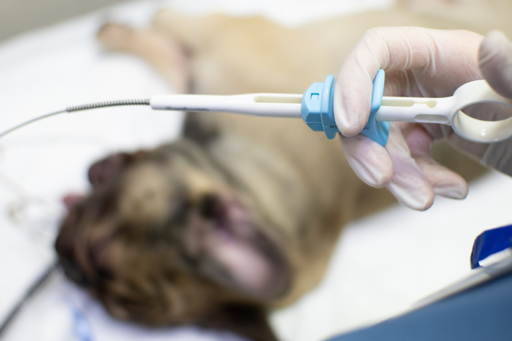
Biopsies come in a few forms: a needle-core biopsy pulls a narrow cylinder of tissue, an incisional biopsy removes a wedge of a larger mass for identification before surgery, and an excisional biopsy takes the whole lump at once, which can serve as both diagnosis and treatment. Needle biopsies can often be done using ultrasound with no or only light sedation, while an incisional and excisional biopsies require surgery under anesthesia.
| Fine needle aspirate | Biopsy | |
| What it samples | Loose, individual cells | An intact piece of tissue |
| Sedation | Usually none | Sedation or anesthesia |
| What it shows | Cell type, often benign versus malignant | Tumor type, grade, invasion, margins |
| Best suited for | A quick first look at accessible lumps | Definitive diagnosis and surgical planning |
| Turnaround | A few business days | Longer, since tissue must be processed |
Which Lumps Skip the Needle Test Altogether?
A few situations bypass the needle test entirely because a scattering of cells simply can’t answer the question. When a mass sits on an organ that bleeds easily, near delicate structures like eyes or blood vessels, or when two very different diseases produce nearly identical cells, tissue architecture is the only reliable path.
Splenic Masses: Harmless Growth or Hemangiosarcoma?
A mass on the spleen is one of the hardest calls in veterinary medicine. Roughly half of splenic tumors are benign and half are malignant, and no ultrasound or radiograph reliably tells which is which. Hemangiosarcoma in dogs is an aggressive cancer of the blood vessel lining that often takes hold in the spleen, where a mass can rupture and bleed heavily before any outward sign appears.
Aspirating the spleen is both unreliable and risky, since the organ is full of blood and a needle can trigger dangerous bleeding. We often recommend removing the whole spleen instead, a procedure called a splenectomy, and sending it for histopathology, so the diagnosis comes after the tissue is safely out. A spleen that ruptures is a true crisis, and our around-the-clock emergency care means a dog who suddenly collapses with internal bleeding can be seen right away, day or night.
Chronic Vomiting in Cats: IBD or Lymphoma?
A cat who has been vomiting for months, dropping weight, and picking at meals presents another puzzle an aspirate can’t solve. Inflammatory bowel disease in cats produces chronic vomiting, diarrhea, and weight loss that look virtually identical to small-cell intestinal lymphoma on an exam, on bloodwork, and on ultrasound.
The only reliable way to tell them apart is a full-thickness biopsy of the intestinal wall, which captures how the cells are organized through every layer. That distinction matters enormously: one condition is managed with diet and anti-inflammatory medication, while the other calls for a very different treatment path.
What Happens After Your Pet’s Lump Results Come Back?
Results generally land in one of three buckets, each with a different next step.
A benign result means the mass isn’t cancer, and the plan is usually simple: keep an eye on it and mention any change at your pet’s next visit.
A malignant result means the choices depend on the tumor type, its grade, and whether it has spread. Removing the mass, staging tests like x-ray and ultrasound to check for spread, and a referral for oncology input are all on the table, and we’ll map out the best plan for your pet with you. When comfort is the priority, our acupuncture and other complementary therapies work alongside gentle pain control to help a pet feel good for as long as possible.
An inconclusive result is more common than you might expect, and it simply means the sample didn’t give a clear enough answer, not that you’ve run out of options. Further diagnostic testing, whether repeating the aspirate, moving up to a biopsy, or adding imaging, usually gets us to a confident diagnosis.
How Do You Keep an Eye on a Benign Lump at Home?
A benign result doesn’t close the book, because new masses can form alongside the one we already checked, and a few lump types can change over time. Simple home monitoring, tracking size, shape, and feel, catches those changes early and tells us when a recheck is worth scheduling.
A monthly habit works well: during a cuddle or grooming session, run your hands over your pet and note each lump’s location, rough size, and how it feels. Around Saylorsburg, a heavy winter coat can hide a new bump for months, so hands find what eyes miss. Some families snap a phone photo next to a coin or jot a quick note, so a slow change doesn’t slip by unnoticed. Here’s what should prompt a call rather than a wait-and-see.
| Change you notice | Why it’s worth a call |
| Rapid growth over days or weeks | Fast change can signal a more active tumor |
| A soft lump turning firm or anchored to deeper tissue | Suggests the mass may be invading rather than just sitting on top |
| Redness, heat, discharge, or an open sore | Points to infection, inflammation, or an ulcerating growth |
| Licking, limping, or flinching near the spot | The mass may be painful or pressing on something |
Even a confirmed lipoma may eventually need removal, not because it turned dangerous, but because a large fatty mass in an armpit or behind a leg can rub, get in the way, or limit how comfortably your pet moves. When something on your tracking list shifts, regular recheck exams let us compare it against the baseline and decide whether to resample.
Frequently Asked Questions About Aspirates and Biopsies for Pet Lumps
Is a fine needle aspirate safe for my pet?
For most skin lumps, a fine needle aspirate is very safe and about as involved as a vaccination. Because sedation is rarely needed, there’s no anesthetic risk to weigh. The main exceptions are masses near delicate structures or organs that bleed easily, like the spleen, where we choose a different, safer approach.
Why can’t you just remove the lump and skip the testing?
Sometimes removing the whole mass is the right first move, especially for a small, accessible one. But sampling first often changes how the surgery is done. Knowing a tumor is a mast cell tumor, for example, tells us to take wider margins the first time, which gives your pet the best shot at a complete removal. Cutting blind can mean a second surgery if the margins come back dirty.
How long does it take to get results back?
A fine needle aspirate read as cytology usually comes back within a few business days, sometimes sooner. A biopsy takes longer, because the tissue has to be processed, sliced thin, stained, and examined by a pathologist, which often means several days to a week or more. The wait can feel endless when you’re anxious, so reach out any time with questions while results are pending. We’d rather answer a small worry than have you sit with it.
Getting Your Pet’s Lump Checked Sooner Rather Than Later
The sooner a new lump is tested, the more choices you and your pet have. A mass that turns out to be a harmless fatty deposit hands you peace of mind, and one that turns out to be serious is far more treatable when it’s caught small. If you’ve found something new on your dog or cat, schedule a visit with Creature Comforts Veterinary Service in Saylorsburg and we’ll get you a real answer.